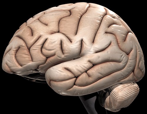
How the brain handles words
 Washington, Apr 30 : How the brain gives meaning to letters on a page has been a mystery for scientists. Now, a new study has tried to solve the puzzle.
Neuroscientists at Georgetown University Medical Center have found that an area known to be important for reading in the left visual cortex contains neurons that are specialized to process written words as whole word units.
Although some theories of reading as well as neuropsychological and experimental data have argued for the existence of a neural representation for whole written real words (an "orthographic lexicon"), evidence for this has been elusive.
"Reading relies on neural representations that are experience dependent," says senior author Maximilian Riesenhuber, PhD, of the GUMC Laboratory for Computational Cognitive Neuroscience.
"Evolution did not provide each of us with a little dictionary in our heads," the expert added.
Because the findings, published in the April 30 issue of Neuron, shed light on how written words are processed in the brain, they also provide clues as to how reading disorders such as dyslexia could arise, Riesenhuber says.
"Previous studies have shown that this brain area is affected in reading disorders such as dyslexia, but it is unclear what the mechanisms involved are. Our data suggest that looking at the neuronal selectivity in this area might provide new insight. For instance, we would expect reading difficulties if neurons never become well tuned to words, making reading a slow, arduous process, just like it would be if reading all nonwords," the expert added.
The GUMC researchers - Riesenhuber, first author Laurie S. Glezer, MA, and Xiong Jiang, PhD - set up a series of experiments with the participation of volunteers. They showed the participants pairings of words, and used functional Magnetic Resonance Imaging (fMRI) to measure brain blood flow in an area in the left visual cortex called the "visual word form area" while the participants performed a reading task.
Most other studies using fMRI to examine the "visual word form area" have used the averaged neuronal response in which many word stimuli are presented and the change in activity is measured, but this approach does not tease out the response neurons have to individual words, Riesenhuber says. However, by using the technique of fMRI rapid adaptation, in which the stimuli are shown in pairs, it is possible to measure the selectivity of neurons for individual words.
In their experiments, the researchers looked at the response between two visually similar normal words that shared all letters but one (i. e. ''boat'' and ''coat'') and found that the neural response to this condition "looked just like when participants saw two words that shared no letters, for example ''coat'' and ''fish''," says Glezer.
"This shows that the neurons in this area of the brain are very selective for individual words. Even though the two words shared all letters but one, there is no overlap in the neural representation, just like when the two words are completely different," the expert said.
The researchers then looked at the brain''s response to sets of nonwords in which the stimuli look like real words but have never been seen before (i. e. tarm). They found that the response to nonwords was not selective, with similar nonwords appearing to have overlapping neural representations. (ANI)