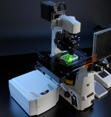
Novel laser microscope can diagnose skin cancer in single snapshot
 Washington, Aug 30 : Physicists from the Consiglio Nazionale delle Ricerche (CNR) in Rome have developed a new type of laser scanning confocal microscope (LSCM), which could diagnose skin cancer in a single snapshot.
Typical LSCMs take 3-D images of thick tissue samples by visualizing thin slices within that tissue one layer at a time.
Sometimes scientists supplement these microscopes with spectrographs, which are devices that measure the pattern of wavelengths, or "colours", in the light reflected off of a piece of tissue.
This pattern of wavelengths acts like a fingerprint, which scientists can use to identify a particular substance within the sample.
But the range of wavelengths used so far with these devices has been narrow, limiting their uses.
The new microscope has overcome this problem.
Unlike other combination "confocal microscope plus spectrograph" devices, the new machine is able to gather the spectrographic information from every point in a sample, at a wide range of wavelengths, and in a single scan.
To achieve this, the authors illuminate the sample with multiple colours of laser light at once - a sort of "laser rainbow" - that includes visible light as well as infrared.
This allows scientists to gather a full range of information about the wavelengths of light reflected off of every point within the sample.
Using this method, the researchers took high-resolution pictures of the edge of a silicon wafer and of metallic letters painted onto a piece of silicon less than half a millimetre wide.
They also demonstrated that it is possible to apply this technique to a tissue sample (in this case, chicken skin) without destroying it.
With further testing, the researchers say the microscope could be used to detect early signs of melanoma; until then, it may be useful for non-medical applications, such as inspecting the surface of semiconductors.
The discovery has been described in a paper accepted to the AIP's new journal AIP Advances. (ANI)