3-D printed tumor cells will imitate cancer cells to aid research
 In a recent development scientists have effectively developed a 3D model of a carcinogenic tumour with the help of a 3D printer. This new technique can now be employed to test the viability and safety of new cancer medications and treatments.
The model comprises of a network structure, 10 mm in width and length, composed of gelatin, alginate and fibrin, reproducing the fibrous proteins which the extracellular grid of a tumours are made up of.
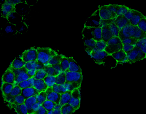
The model comprising of a framework of proteins made with fibre covered in cervical tumour cells, has given a reasonable 3D representation of the surroundings of cancer cells which could help in the finding of new medications and cast new light on how cancers are initiated, developed and end up spreading all around the body.
The network structure is covered in Hela cells which are special, undying cell line that was initially inferred from a cervical malignancy persistent in 1951. In spite of the fact that the best method for contemplating cancer cells is to study them via clinical trials but limitations of safety and ethical nature make it troublesome for these sorts of studies to be done largely.